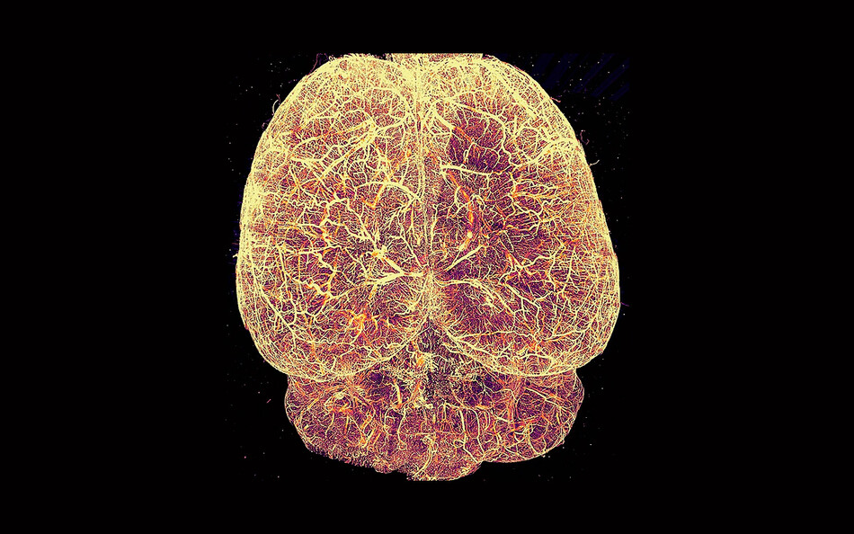
A team of researchers led by Columbia biologist Raju Tomer has developed a powerful, affordable, and user-friendly microscope that could dramatically increase the number of scientists around the world who are able to conduct cutting-edge biomedical research. The new technology, a type of light-sheet fluorescence microscope (LSFM), produces highly detailed three-dimensional images of living tissue. It is much cheaper and easier to use than existing LSFM tools, Tomer says, which could expand access to 3D imaging.
“Similar microscopes sell for upwards of $700,000 and are difficult to operate and maintain, so they tend to be available only in advanced imaging facilities at major universities,” he says. “Ours costs one-tenth as much and is largely automated.”
Tomer and his colleagues, who describe their invention in a recent issue of the journal Nature Biomedical Engineering, lowered costs by swapping out ultra-expensive components typically found in light-sheet microscopes for off-the-shelf alternatives. “For our light source, instead of a $40,000 laser apparatus, we used a $300 version found in ordinary video projectors,” Tomer says. “We also incorporated simpler optics and electronics, and the finished product is even more robust.”
The Columbia team has partnered with the Vermont-based technology company MBF Bioscience to bring their microscope to market, while also sharing the design details online, allowing scientific institutions to build the tool themselves.
Tomer says the new tool could be used for any number of research applications, from observing the impact of novel drugs on human organoids in petri dishes to mapping the interactions of brain cells for clues about what goes wrong in neurological conditions. He notes that the tool could also accelerate the development of new medical diagnostics. By making high-quality 3D imaging more widely accessible, especially in middle- and low-income countries where advanced microscopes are in short supply, he says, the technology could help scientists process the huge numbers of tissue samples that are needed to train AI models to detect diseases like cancer.
“And if AI models are trained on samples from all over the world, they will be less biased, and more accurate, for everyone,” he says.