Pharmaceutical companies today are spending millions of dollars developing fluorescent dyes that can be injected into mice and then made visible by shining an ordinary light through the animal, just as a child might peer into his hand with the help of a flashlight. Dyes that glom onto cancer cells, for instance, can reveal the size of a tumor, enabling researchers to monitor how it responds to medication. The imaging procedures are designed specifically for tiny lab animals, whose bodies are easily illuminated, so visible light does the trick - x-ray and MRI technology typically isn't necessary.
A shortcoming of in vivo imaging techniques, however, is that the light that finds the dye shines right through internal organs, making them invisible even to the most highly sensitive cameras. "That makes it difficult to know exactly where the tumor is in relation to organs," says Columbia biomedical engineering assistant professor Elizabeth Hillman, "especially because organs will shift slightly as the animals grow."
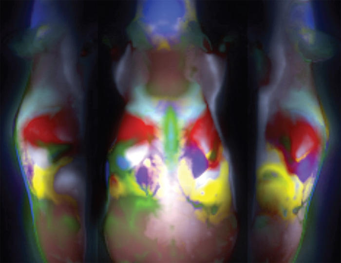
Hillman thinks she's solved this dilemma. Whereas current in vivo imaging techniques involve taking a single photograph to show where fluorescent dye eventually accumulates in an animal, Hillman has invented a way to record movies of the dye circulating throughout the animal. She says that the dye enters various organs at slightly different times, but that all parts of the liver, for example, fill up with dye together. By analyzing each pixel of every frame in her movie to determine when individual pixels show the presence of dye, Hillman can map the borders of every organ. She then creates color-coded images showing their locations.
"This will give researchers very cheap and simple anatomical overlays to reveal whether a tumor is touching the liver, the kidney, the gut, or all of them," says Hillman. Her technology also could generate more definitive images of tumors. "Using current imaging techniques, the movement of dye in the animal's body often creates noise in results because some of the dye might not have accumulated in the tumor when the photograph is taken," she says. "By showing where the dye is moving, and in what tissues it is increasing, we can provide an extra level of information."
Hillman's study appeared in the September issue of the journal Nature Photonics.