By the time they landed at Kennedy International Airport on the morning of January 26, 2015, nine-year-old Myrrah Shapoo was already suspicious. Her father had promised her the trip would be fun — that they would sit in the fancy seats at the front of the plane and watch cartoons. But her mother had been crying as she rushed to pack their suitcases, and it seemed strange that all her aunts and uncles had suddenly shown up at the family’s New Delhi home to hug and kiss Myrrah goodbye.
Now Myrrah was wondering why her father insisted on pushing her through the airport terminal in a wheelchair when, really, she could have walked. And why did she have to wear a silly paper mask? Finally, when a distant relative greeted them outside the baggage claim and asked her father in hushed tones about “her condition,” Myrrah figured it out.
“Papa,” she said, “am I sick again?”
In the summer of 2012, after countless doctor’s appointments, dozens of tests, and months spent watching their six-year-old daughter suffer from unexplained fevers, achy bones, and deep and persistent tiredness, Sajid and Rubina Shapoo got the awful diagnosis. Their daughter Myrrah had acute lymphoblastic leukemia (ALL).
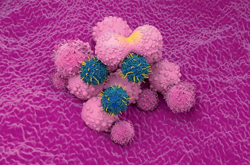
ALL is a cancer of the blood — specifically of the white blood cells. Typically those cells work with the immune system to fight infection and protect the body against disease, but in children with ALL, an uncontrollable proliferation of cancerous white blood cells upsets the delicate constitution of the bloodstream, crowding out normal white blood cells, as well as red blood cells and platelets. This makes it impossible for the body to properly fight infection, absorb oxygen, or heal wounds.
Acute lymphoblastic leukemia is the most common form of cancer in children. It is also one of the most treatable; nearly 90 percent of children diagnosed with the condition are cured. Myrrah’s parents, after getting over their initial shock, were optimistic that she would survive.
“The doctors told us that if you had to get cancer, this was the one with the best prognosis,” says Sajid, who works as a police official in India. “And, clearly, when you’re told that your daughter has a disease that most kids recover from, you expect her to be among the fortunate ones.”
Myrrah was initially treated at New Delhi’s Indraprastha Apollo Hospital, which boasts a state-of-the-art cancer clinic, considered among the best in India. Her doctors there followed the same diagnostic and treatment protocols that physicians at most hospitals in the US or Europe recommend. Within hours of determining that her white blood cells were cancerous, they sent a sample of Myrrah’s blood to a laboratory for karyotyping, a form of genetic analysis that involves examining a cell’s nucleus through a microscope to see if any of its forty-six chromosomes are missing or misshapen. The scientists who examined Myrrah’s blood were looking at those X-shaped bundles of coiled-up DNA for clues. For instance, if a particular section of chromosome 9 was lopped off and fused onto the end of chromosome 22, it would indicate a mutation commonly called the “Philadelphia chromosome.” This would suggest that Myrrah had a subtype of leukemia that is treatable with Gleevec, one of a handful of cancer drugs that have been designed to target specific genetic aberrations. But there was no Philadelphia chromosome and no other hints as to what was driving Myrrah’s illness.

Without a clear road map for treatment, her doctors did what any other oncologists would do in the situation: they prescribed a cocktail of four common chemotherapy drugs — dexamethasone, vincristine, daunomycin, and PEG-asparaginase — to be administered every day for a month through a port that would be implanted in Myrrah’s neck. The physicians at Apollo Hospital would subsequently give her several additional chemotherapy cocktails — typically administered for a month or so at a time — in order to maximize the chances that any cancer cells that withstood one drug would be destroyed by another. If leukemia cells migrated to her spinal column or brain, where chemotherapy drugs couldn’t penetrate the blood-brain barrier, they would also bombard Myrrah with radiation.
To understand the suffering that Myrrah would go through in those first arduous months of treatment, it helps to know how chemotherapy works. These drugs, most of which were developed in the 1940s, ’50s, and ’60s, an era of aggressive chemical experimentation on human cancer patients, attack various molecules in the body that are necessary for cell growth and cell division. The idea is that since cancer cells grow and divide at a maniacal pace, they are preferentially affected and die off before the rest of the body breaks down. But these drugs take a severe toll on all cells that divide rapidly. They kill those found in hair follicles, which makes patients go bald; they attack the lining of the digestive tract, which causes severe nausea and diarrhea; and they annihilate the mucous membranes, which can lead to painful sores in the mouth and on the lips. Myrrah experienced all of these side effects and more: the drugs made her mentally foggy and weakened her immune system so badly that she developed repeated infections; on one occasion, she nearly died of meningitis. During this period Myrrah lived at the hospital for weeks at a time, and her parents took turns sleeping next to her on a cot. Doctors stopped by almost every day bearing needles — small needles for delivering shots, medium-sized needles for taking blood, and impossibly long, thick needles for drawing spinal fluid out of her back, which made her scream in pain.
“We nicknamed her the Cancer Slayer because she is such a fighter,” says her father. “Sometimes she would ask me, ‘Why don’t other kids have to do this?’ And I would say, ‘Because only you are strong enough. Only you can take it.’”
And she did take it. Little by little, she got better. By the spring of 2013, there was no more cancer detectable in her body, and doctors stopped the most powerful chemotherapy regimens. They would continue to give her relatively mild doses of chemotherapy for the next two years, just in case any traces of cancer still lingered, but Myrrah soon regained her strength and returned to school. That summer, her parents celebrated by taking her on vacation to Europe. Life was, for a few precious months, normal again.
Then, in January 2015, during a routine checkup, Sajid and Rubina learned that Myrrah’s cancer had returned. This time, the doctors said there was nothing more they could do. “I almost blacked out,” says Sajid. “I couldn’t see anything, and there was this loud buzzing in my head that drowned almost everything out. After a few seconds, I heard my voice saying: ‘No, this cannot be true.’”
The doctors’ only suggestion was that the Shapoos take their daughter to the United States — specifically, to New York–Presbyterian/Columbia University Medical Center — where a group of pediatric oncologists was developing a new, personalized approach to treating cancer by conducting an extremely detailed analysis of each patient’s DNA. “We were told that Myrrah’s disease might somehow be treatable, but that it would require an unusually in-depth investigation to figure out how,” says Rubina, who is a television journalist and documentary filmmaker. “This was our only chance.”
Two days later, on a Friday afternoon, Sajid requested a long-term leave from his job and booked two tickets to New York. He and Myrrah left that weekend. His wife would remain in New Delhi with their newborn son, Hayder, and Myrrah’s twelve-year-old brother, Ruhayl.
“This was the darkest hour for me,” says Sajid. “I’d always been the one in our family refusing to entertain bad thoughts about Myrrah’s prognosis. But now I struggled to keep myself together for my wife and for my little girl.”
New Terrain
In 2014, the pediatric oncology unit at Columbia University Medical Center became one of the first in the world to offer whole-exome sequencing to every child with cancer in its care. Whole-exome sequencing is a method of DNA profiling that reveals the exact sequence of all thirty million nucleotides — adenine, thymine, guanine, and cytosine molecules — that constitute a person’s nineteen thousand genes.
Because using this technology is expensive and time-consuming, and because it generates an overwhelming amount of data, physicians rarely use whole-exome sequencing as a diagnostic tool. Typically, oncologists simply want to know whether the person has any of a handful of genetic mutations whose roles in cancer are well understood. This is accomplished with karyotyping (the method that scientists at Apollo Hospital had used) and with slightly more sensitive computer-based genetic tests that scan a person’s DNA for limited numbers of mutations that have been proven to cause cancer.
Columbia’s pediatric oncologists, however, sought to look more comprehensively across all genes, with the hope of discovering ways they might save children with forms of cancer that were deemed incurable. Working alongside data scientists, cell biologists, molecular pathologists, and other medical researchers, they were determined to embrace whole-exome sequencing in all its complexity.
“By looking not only at the genes whose contributions to cancer are already well established for a given condition, but at the countless others that researchers have identified as potentially being involved in other forms of cancer, we knew we could gain an unprecedented level of insight into each child’s disease,” says Andrew Kung, the chief of pediatric hematology, oncology, and stem-cell transplantation at CUMC. “If a child didn’t respond to the standard treatment regimen, we’d have all the information we could possibly want at our fingertips when considering alternative approaches.”
Kung envisioned a network of clinicians and researchers working side by side, seamlessly integrating information the researchers gleaned from whole-exome sequencing into treatment decisions on a daily basis. He dubbed his new program Precision in Pediatric Sequencing (PIPseq).
When Kung launched PIPseq with the support of Lee Goldman, the chief executive of CUMC, and Stephen Emerson, the director of CUMC’s Herbert Irving Comprehensive Cancer Center, he knew it would be a gamble. Assessing the significance of the countless possible mutations scattered across a single person’s nineteen thousand genes was an ambitious task, usually undertaken only as part of a major research project aimed at identifying genes associated with a particular disease. In clinical situations, whole-exome sequencing was used sparingly. Health-insurance companies almost never paid for it, and few studies had even attempted to quantify the potential clinical payoff of analyzing DNA on this scale.
“It was a chicken-or-egg dilemma,” says Kung. “Since no hospital had ever made a serious attempt to show what comprehensive sequencing could achieve in the clinic if used routinely, its benefits were unproven.”
But Kung, who had been recruited to Columbia from Harvard in 2012 in part to advance the use of genomics in pediatric oncology, believed that his division had an opportunity to help shape the future of medicine. It was only a matter of time, he was convinced, before this type of highly sophisticated genetic sequencing became cheap enough, fast enough, and easy enough to use routinely, not only for the diagnosis and treatment of cancer but for a wide variety of conditions. He saw that research on human genetics was already having profound intellectual implications, causing scientists to rethink the definitions of many diseases. It had revealed, for example, how cancers that started in different parts of the body sometimes shared the same physiological roots and were responsive to the same medicines. He anticipated that genetics research would also have disruptive effects on how medical institutions are organized. Gone would be the days when physicians operated more or less independently from laboratory scientists, learning about medical breakthroughs at annual conferences and then going back to their clinics to ponder the findings. He envisioned a hospital in which physicians would be in constant conversation with geneticists, cellular biologists, and molecular pathologists. Kung thought that CUMC, which was making major investments in its genomics research and clinical programs under the umbrella of its recently established Institute for Genomic Medicine, was ideally positioned to figure out how these new types of collaborations should work. His bosses agreed, and in the fall of 2014, Columbia University Medical Center began offering DNA sequencing and analysis to the family of every pediatric cancer patient, on the understanding that CUMC would pick up the estimated $8,000 tab if the family’s insurance company declined to pay.
“I think that many other institutions were waiting for genetic sequencing to become cheaper and easier,” says Kung. “My colleagues and I thought that CUMC should chart the way forward, even if we had to absorb the costs.”
Eleventh Hour
When a person with advanced leukemia has blood drawn, the fluid that comes out is thin, watery, and pinkish. This is because the spongy tissue inside the person’s bone marrow, which is where all blood cells are manufactured, is now devoting itself almost exclusively to producing monstrously deformed cancerous white blood cells.
By the time Myrrah Shapoo arrived at CUMC in January 2015, her blood had a ghostly translucence. She was also beginning to feel lethargic, the first sign of oxygen deprivation. Without treatment, she likely would die within days or weeks — either from infection or from her oxygen-starved organs shutting down.
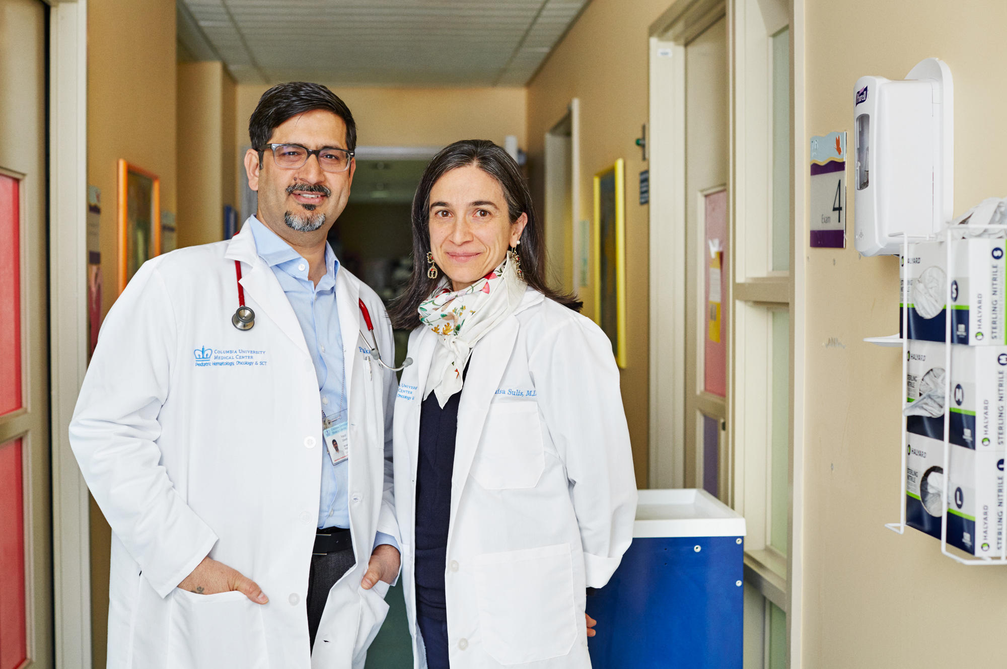
To stabilize her, a team of physicians led by Maria Luisa Sulis ’15PH, a childhood-leukemia specialist, opted to give her a cocktail of chemotherapy drugs similar to the one she had received during her first few months of treatment in India. Based on the course that Myrrah’s disease had taken, they could tell that those drugs had done a decent job of combating her cancer.
“The problem seemed to have occurred later, with the drug she was given at the end of her treatment, after her cancer levels had dropped so low as to be undetectable,” says Sulis. “Our goal now was to knock her cancer cells back down to that level and keep her alive long enough for the genetic analysts to help us figure out a more personalized path to take.”
Soon after Sajid and Myrrah arrived at Columbia, the rest of the family joined them in the United States. They stayed with cousins in New Jersey and would make the long commute to the intensive-care unit of CUMC’s pediatric cancer ward every day. When Myrrah was strong, the family would Skype with relatives back home. When Myrrah felt too ill to socialize, Sajid or Rubina would read her stories from a collection of Indian adventure tales or simply cuddle with her on her tiny bed.
“Back in New Jersey each night, I’d bury myself in scientific papers about the genetics of leukemia,” says Sajid, a handsome and gentle-natured man of forty-one. “It was important to me that I try to understand what was going on with my daughter’s disease. Somehow that made the situation more tolerable.”
As Myrrah fought for her life, clinicians and scientists from several CUMC departments and research laboratories joined forces to figure out a treatment plan. First on the case were pathologists at CUMC’s Laboratory of Personalized Genomic Medicine, a diagnostic facility that provides genetic testing for a wide range of diseases. They had received a vial of Myrrah’s blood and bone-marrow tissue the day after she arrived at the hospital and had immediately gone to work extracting DNA from both her cancerous white blood cells and her healthy cells. After processing the DNA, they loaded it onto a glass slide, which they placed into a computerized sequencing machine. The sequencer, which painstakingly examined strands of DNA to determine the identity of all thirty million nucleotides in the protein-coding genes of each cell, then downloaded two composite genetic blueprints: one representing a typical cancer cell in Myrrah’s bloodstream, and the other its healthy counterpart. Next, software designed by bioinformaticians in the laboratory organized that data into spreadsheets that highlighted the differences between the two. This would enable scientists to spot not only mutations in Myrrah’s cancer cells but also differences between the DNA in Myrrah’s noncancerous cells and the DNA of an average human cell — as represented by the findings of the Human Genome Project.
“What comes next is a massive cross-referencing project, in which you’re searching for commonalities among the genetic mutations you find in the patient and those that have been documented as possibly contributing to leukemia,” says Mahesh Mansukhani, an associate professor of clinical pathology who, as director of the Laboratory of Personalized Genomic Medicine, oversees the genetic sequencing and analysis done as part of PIPseq. “Your analysis goes in both directions — you see if the patient carries any mutations described in the scientific literature, and you scan the literature for references to variations that the child is carrying.”
Mansukhani and other members of his lab began analyzing Myrrah’s genetic profile three days after she was admitted. Over the next several weeks, they would consult regularly with pathologists, data specialists, and oncologists from other CUMC labs to help them determine which statistical correlations were most relevant. This is crucial, Mansukhani says, because a team of analysts could easily spend years searching for meaningful patterns in data sets as vast as those his team wrestles with.
“Consider that you’re looking at nineteen thousand plus genes in this child, and that fifteen or twenty genetic aberrations have so far been definitely linked to acute lymphoblastic leukemia,” says Mansukhani. “That doesn’t sound like an impossible puzzle to solve, right? But consider that each of those genes linked to leukemia has numerous mutated forms, each of which may have a different physiological effect. And consider that dozens of other genetic errors have been hypothesized to contribute to leukemia in small numbers of kids who have rare subtypes of the disease. Now it’s becoming a very complex mathematical problem, right? Meanwhile, you’ve got a child whose family is depending on you to solve this puzzle pretty darn quickly. So you’d better have a plan about what you’re looking for when you turn on your computer.”
One Girl’s Illness, Decoded
By early February 2015, a picture began to emerge of what was making Myrrah’s disease so stubborn. The first breakthrough occurred when Susan Hsiao, a molecular pathologist in Mansukhani’s group, discovered that Myrrah’s cancer cells carried a mutation in a gene called NT5C2. This mutated gene does not cause cancer, but it can derail a patient’s treatment by making leukemia cells resistant to certain chemotherapy drugs, including 6-mercaptopurine, or 6-MP. One of the first chemotherapy drugs ever invented, 6-MP is often given to leukemia patients in the very last stage of treatment in what is called the “maintenance” period of chemotherapy. Myrrah had received it every day for nearly two years, from the spring of 2013 through the end of 2014.
“She had variants in others genes linked to cancer, too, but this one stuck out as being the most immediately relevant to her care,” says Hsiao. “It provided a perfect explanation for why she had relapsed.”
The mutation that Hsiao found in Myrrah’s NT5C2 gene was the simplest type of genetic aberration there is: a point mutation, which is the result of a single nucleotide being missing, inserted where it doesn’t belong, or swapped out for another. In Myrrah’s version of NT5C2, the 1,219th of its 1,683 nucleotides, which should have been a guanine molecule, was a thymine molecule.
Hsiao says that in the course of her analysis she will sometimes contact scientists who have studied a particular gene to discuss the clinical ramifications of the variation she has found. In this case, her job was easier than usual: the scientist who had first described NT5C2’s role in chemotherapy resistance was a CUMC leukemia researcher named Adolfo Ferrando. Ferrando is an expert on the genetic and molecular basis of the disease, and in 2013, his research team had analyzed the genes of 140 leukemia patients who had relapsed after receiving 6-MP or a closely related drug. The scientists had found that 15 percent of the patients had mutated versions of NT5C2.
“Adolfo’s team was able to confirm that a point mutation at that location was likely to cause problems,” says Hsiao.
Now Myrrah’s physicians knew what drug not to give her. But what should they give her instead? To answer this question, the PIPseq team turned again to Ferrando, whose laboratory is among several at CUMC that regularly provide analytic support to the pediatric oncologists. Ferrando’s team, in addition to analyzing the chemical composition of DNA, can analyze the RNA and proteins made by genes and determine which genes are active and which are inactive, both in normal development and in cancer.
“Genes are not static entities,” says Ferrando. “They turn on and off, often as a result of their interactions with other genes. And sometimes you need that level of understanding to tease out what’s happening in a particular person’s disease.”
In April, Alberto Ambesi-Impiombato, a postdoctoral researcher in Ferrando’s group, made an important discovery. Using a computational technique called cluster analysis, he observed that hundreds of genes in Myrrah’s cancer cells were turning on and off in a pattern that suggested her disease shared deep similarities with the form of ALL that is caused when a part of chromosome 9 fuses onto chromosome 22, resulting in what is known as the Philadelphia chromosome. The drug Gleevec was designed specifically to treat that form of ALL; it works by dismantling a mutant protein produced by the Philadelphia chromosome that tells other molecules inside of a cell to continually make the cell divide.
“So now the question was whether or not this patient’s disease was driven by a similar mechanism,” says Ferrando. “And if it was, perhaps Gleevec or a similar drug might be used therapeutically.”
Maria Luisa Sulis, the physician overseeing Myrrah’s care and herself a former laboratory scientist, solved the final piece of the puzzle. She ordered targeted DNA tests to look for a handful of genetic mutations that are known to cause malfunctions in a cell’s metabolic signaling very similar to those caused by the Philadelphia chromosome. And they found one: in Myrrah’s cancer cells, one entire gene in chromosome 9 had been folded back onto its neighbor so that their nucleotides mixed to create a hybrid gene known as NUP214-ABL1. The protein manufactured by this hybrid gene had previously been shown to act similarly to the one produced by the Philadelphia chromosome, instructing a cell to incessantly divide.
“This was a chromosomal rearrangement much smaller and more difficult to spot than the Philadelphia chromosome,” says Mansukhani. “You wouldn’t see this one unless you analyzed how the DNA expresses itself as RNA.”
The finding was all the more remarkable because it pointed to a specific treatment strategy. In 2006, the pharmaceutical company Bristol-Myers Squibb released a drug called Sprycel, which works very similarly to Gleevec, targeting the specific types of proteins that both the Philadelphia chromosome and the NUP214-ABL1 gene produce. Experiments had shown that Sprycel would be particularly effective in Myrrah’s case.
So now the path forward was clear: Myrrah’s doctors would add Sprycel to the traditional chemotherapy cocktail she was receiving, and continue giving her the drug for as long as it took to wipe out any remaining cancer cells.
Hope for Others
On a recent Wednesday morning, about twenty-five Columbia physicians and medical researchers gathered in a conference room at CUMC to update one another on cases they were working on as part of the PIPseq program. Mansukhani took the floor first, presenting his team’s analyses of the DNA of several children recently admitted to the hospital. He spoke in the dense, super specialized language of genetics, using alphanumeric code to describe the glitches in the children’s genes. “We found a mutation at c.884C>T, p.P295 in 104 of one thousand cells,” he said, referring to a young boy with a tumor. The physicians peppered him with questions: “Isn’t that in the same region as the famous Sonic Hedgehog mutation?” “Would we expect to find a mutation there in someone with this kind of tumor?” “What does this tell us about his chances of relapse?” Andrew Kung, after presiding over a lengthy discussion of the case, summarized the group’s consensus: the physicians should start the boy on a standard chemotherapy regimen, and the analysts would continue looking for clues about what experimental drugs he might be given, should he need an alternative.
After the meeting, Kung expressed his excitement at seeing physicians and medical researchers collaborating so intimately: “Five years ago, that wouldn’t have happened. It’s our ability to bring together experts from across the entire medical center that makes precision medicine work.”
Since the PIPseq program was launched, in 2014, more than one hundred pediatric cancer patients have had their genes sequenced at CUMC. In about two-thirds of these cases, Kung says, Columbia researchers have discovered something in a child’s DNA or RNA that has helped physicians decide how to treat the patient. The researchers’ discoveries have led to children receiving gene-targeting drugs like Gleevec or Sprycel in just a handful of cases, Kung says, because there are only a few targeted therapies approved for use in children. But the genetic analyses have guided treatment in other ways. On many occasions, they have indicated that a child has a particularly aggressive type of cancer and needs unusually high doses of chemotherapy. In other instances, the analyses have motivated physicians to perform a bone-marrow transplant earlier in the treatment process than is typical.
Conversely, the Columbia researchers’ analyses have sometimes indicated that physicians could save a child with unusually low doses of chemotherapy, or without a bone-marrow transplant. Physicians would prefer to scale back treatment if possible, because all cancer treatments carry risks: the toxicity of chemotherapy drugs, for instance, not only cause severe short-term side effects but also increase a patient’s long-term risk of developing other cancers; and a donor’s bone marrow might be rejected by the recipient’s body, or even attack it, both of which are often fatal.
“One of the most active areas of leukemia research today is figuring out how to identify patients who require less intensive treatment,” says Kung. “Just because we’re curing 90 percent of children with ALL doesn’t mean that we accept the toxicities that we inflict in pursuit of a cure.”
It is too soon to know if any children who have participated in the PIPseq program have been cured as a result of having had their genes sequenced. This will take several years to establish, since cancer patients are generally considered cured only after having been in remission for five years or longer. One thing that can be said for certain, though, is that there are children alive today because of this program. Among them is Myrrah Shapoo. This spring, just fifteen months after she and her father first arrived at the hospital, looking for a miracle, the family received wonderful news. There is no more cancer detectable in her blood. Her physicians are cautiously optimistic that she will stay in remission, because they have examined her blood using a newer, much more sensitive diagnostic test than the one used by her Indian doctors when they initially announced that she was in remission three years ago. Her recovery seems attributable both to Sprycel and to a transplant that CUMC oncologist Prakash Satwani performed last summer, when he replaced Myrrah’s bone marrow with bone marrow from her older brother, Ruhayl. The transplant seems to have given her immune system the boost it needed to annihilate the few leukemia cells that had remained in her body.
“What we can presume is that Myrrah’s immune system originally had some sort of flaw that enabled the cancer to take root and proliferate,” says Satwani, who has been overseeing her care since the procedure. “You do a transplant in the hope that the donor’s marrow is innately stronger. So far, this seems to be the case.”
The Shapoos are now living in Morningside Heights, where Ruhayl is attending high school and Myrrah, now ten, is being tutored at home. Her parents expect the family to remain in New York City for another year or two so that Columbia doctors can continue to monitor their daughter. This suits Myrrah fine. She has become close friends with several other young cancer patients. And she loves American television — especially the cooking shows, which she credits with inspiring her dream of becoming a chef when she grows up.
“Right now, I’m focused mostly on spaghetti, which my papa lets me cook Friday nights,” she says. “He wants me to be a doctor. I might do that, too. But first a chef.”
There is ultimately more than one life at stake in the effort to save Myrrah. Many other children have similar genetic mutations. How many more children with the NUP214-ABL1 gene are out there? It is impossible for scientists to know, but studies have suggested that NUP214-ABL1 is likely to be among the most common mutations carried by the 10 percent of children with ALL who still cannot be saved through traditional chemotherapy. So identifying these children, and treating them with drugs best suited for their individual cases, could have a pronounced impact on ALL survival rates.
“The development of drugs like Gleevec and Sprycel was a big deal, because they happen to target some of the most aggressive and deadly subtypes of leukemia,” says Maria Luisa Sulis. “Kids with the Philadelphia chromosome had a dismal survival rate before Gleevec was invented. And we need more targeted therapies to be invented, of course. But we also need to identify everybody who could benefit from the targeted therapies we do have. And we’re not yet doing that as well as we could.”
Sulis is now working with other members of the PIPseq team to fine-tune their procedures for identifying children with mutations whose effects are similar to those of the Philadelphia chromosome, in hopes of diagnosing them more quickly. “People often talk about curing cancer as if there is a magic pill we’ll find that heals everybody,” she says. “The reality is much messier, slower, and more incremental. Cancer is hundreds of diseases. Right now, we’re trying to cure Myrrah Shapoo’s cancer. Then we’ll find other kids with the same subtype of the disease and treat them the same way. This is how we’ll push the cure rate for pediatric ALL up from 90 to 95 percent, and so on. And this is how we’ll beat all cancers. We will cure them one by one.”