A team of researchers led by Columbia biomedical engineer Elizabeth Hillman has discovered that the infant brain has a surprising ability to endure fluctuations in its oxygen supply — a finding that could enable physicians to more fully interpret fMRI brain scans of children as young as one or two and gain insights into autism and ADHD.
Since the 1990s, fMRI scans, which identify spikes in brain activity by revealing patterns of blood flow, have been used routinely by clinicians to assess the effects of strokes and traumatic brain injuries. In recent years, the technology has even shown promise in diagnosing conditions such as depression and Alzheimer’s. Researchers who have performed fMRI scans on very young children, however, have tended to get odd results. Often, the tests will indicate that only small amounts of blood are flowing to sections of a child’s brain that, based on his or her mental activity at the time, should require lots of oxygen-rich blood to fuel their operation.
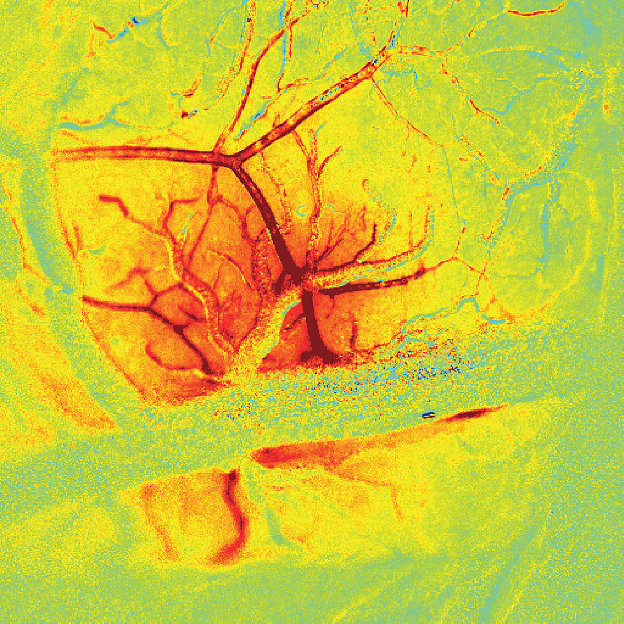
Hillman’s latest research provides an explanation for that phenomenon. In a recent study, her team demonstrated that neurons in the brains of infant mice do not trigger an increase in the flow of blood when firing. Hillman, who is also a principal investigator in the Zuckerman Mind Brain Behavior Institute, hypothesizes that this may be an adaptive trait that helps babies survive the extraordinary transition they make out of the womb.
“All sorts of things can interrupt a baby’s oxygen flow during birth, including the violent squeezing of its body as it comes through the birth canal,” she says. “It makes sense that the brain would be prepared to go without oxygen for a little while.”
Researchers in Hillman’s lab are now preparing to investigate whether the human infant brain behaves similarly to the mouse brain. They are also studying how the brain’s blood vessels grow in response to the oxygen demands of various parts of the organ. The results of this work, they hope, will enable scientists to develop reliable guidelines for interpreting fMRI scans of young children.
“To date, most research on the developing brain has focused on how neuronal circuits take shape,” says Hillman, whose latest paper, written with former PhD student Mariel Kozberg ’15GSAS, ’16PS, appeared in the Journal of Neuroscience. “But we believe that it’s crucial to understand how the brain’s vascular system assembles in tandem with neuronal networks. Recognizing when the brain’s vascular system isn’t developing properly could give us insights into conditions which today truly mystify us.”