While COVID-19 halted most activity on Columbia’s campuses in the spring and summer, it did not stop the march of progress. At the Jerome L. Greene Science Center, at 129th Street and Broadway, scientists welcomed the arrival of a microscope of unmatched capabilities — one that might help unravel some of the brain’s most consequential mysteries.
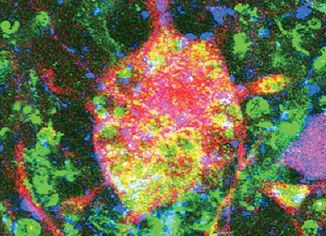
The microscope, which measures two feet wide by three feet high, is called an Aquilos Cryo-FIB: “cryo” because it freezes tissue samples at -274°F and “FIB” because it uses a focused ion beam to peel away cell layers. The refined layer is called a lamella — a window into the cell itself, thin enough to allow electrons to pass through it. And it’s through this window that Anthony Fitzpatrick, an assistant professor of biochemistry and molecular biophysics who runs a lab at Columbia’s Zuckerman Institute, captures images of the tangled proteins that are implicated in neurodegenerative diseases like Alzheimer’s, Parkinson’s, and Lewy body dementia. (See “Toxic Alzheimer’s Proteins Exposed in New Detail” in the Spring/Summer 2020 issue.)
The Aquilos is a delicate instrument. Made by Thermo Fisher Scientific of Waltham, Massachusetts, it is highly sensitive to humidity, electromagnetic fields (areas of energy associated with electrical power), and high- and low-frequency sounds. With the bone-shaking rattle of the elevated 1 train less than a block away and the ongoing deep-excavation construction of the Manhattanville campus, protecting the Aquilos from invisible forces is no small feat: even footfalls can disturb the machine’s equilibrium.
When you consider the microscope’s needs, it becomes clear that it takes many people apart from scientists to make it possible to peer inside a neuron. In a complex renovation overseen by Marcelo Velez ’00BUS, vice president for the University’s Manhattanville development project, a small army of civil engineers, builders, and technicians were tasked with transforming a small room into a veritable fortress for the new machine.
First, Columbia Facilities identified an underused MRI workshop in the bowels of the Greene Science Center, sixty feet below ground level, as far removed as possible from noise inside and outside the building. Next, the walls, floor, and ceiling were coated with epoxy paint to create an anti-moisture “vapor barrier.” A vibration-isolation table, a device containing steel springs and sensors, holds the microscope and keeps it stabilized. And because electromagnetic activity, such as that from the high-voltage lines that run under Broadway, can deflect the microscope’s electron beams and distort its images, workers also installed a magnetic-field cancellation system, which reads the ambient field and produces an opposite, neutralizing field.
“The vibration levels that we measured before we started were borderline, so we considered not taking some of these extraordinary measures,” says Velez. “But in the end, considering that the impact of future construction is unknown, we decided that this was the best strategy to take.”
The acquisition of the Aquilos is part of a $2.84 million National Institutes of Health grant that will allow Fitzpatrick and his team to combine the unrivaled imaging capabilities of the Aquilos with those of the Krios Cryo Transmission Electron Microscope. Also made by Thermo Fisher, the Krios employs technology developed by Columbia biophysicist Joachim Frank, for which Frank won the 2017 Nobel Prize in Chemistry. After Fitzpatrick and his team use the Aquilos to prepare the lamella, they take the sample across the hall to the Krios, which produces three-dimensional images of the proteins by firing electrons into the cell interior. Then the Fitzpatrick lab painstakingly assembles those images on computers to reconstruct the interior of the cell and form a coherent picture of the proteins.
With the Aquilos now firmly ensconced in its specially constructed chamber and ready for action, Fitzpatrick has a dream setup that will allow him to map the structure of the proteins that account for different neurodegenerative diseases. “If you don’t understand how the protein is put together, you can’t figure out how to disassemble and neutralize it,” Fitzpatrick says. “And the Aquilos microscope, paired with the Krios microscope, is the most powerful way of looking at cells that we have.”
This article appears in the Fall 2020 print edition of Columbia Magazine with the title "New Digs for a New Microscope."


