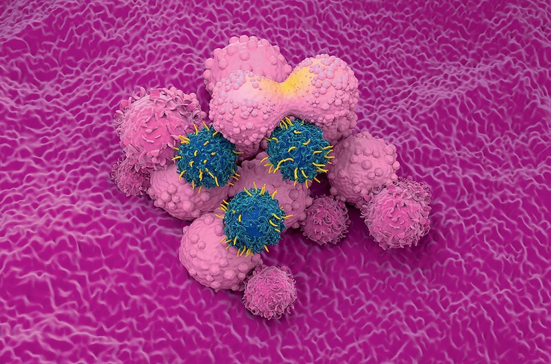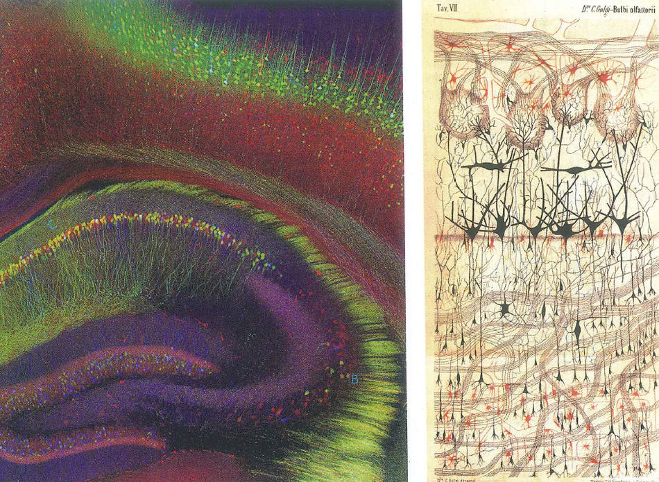
I used to carry images of the brain in my wallet,” says Carl Schoonover ’11GSAS, a fourth-year graduate student in Columbia’s neuroscience program. “I’d show them to other people and their response was, ‘Whoa, that’s the human brain? How is that possible? It’s so beautiful.’ I was surprised that people who didn’t know the meaning of the pictures also found them beautiful. That convinced me that there was interesting material here, and not just for the scientist in me.”
Portraits of the Mind: Visualizing the Brain from Antiquity to the 21st Century (published by Abrams) is Schoonover’s coffee-table tour of how philosophers and scientists have represented that most mysterious of human workings. Schoonover compiled images from archives and from neuroscientists around the world to create a visually explosive book that includes essays by Jonah Lehrer ’03CC, Columbia professors Michael Goldberg and Joy Hirsch, and other leaders in the field. The pictures — from speculative drawings to micrographs to computer-assisted scans — are elegantly simple here, impossibly complicated there.
“Neuroscience is a field with a lot of beautiful data,” says Schoonover, who works in assistant professor Randy Bruno’s CUMC laboratory studying how the somatosensory cortex of rodents processes information from their whiskers to make sense of their environment.
“The selection of images for the book was based on aesthetic value and their having a story to tell. I think the book works because the pictures are beautiful enough that one wants to know what they’re about and how they were made.”